Closed 1/14/10
Totaled 1/10/10 Mr F
Totaled 1/5 /10 Mr F
Totaled 12/22 Mr F
The Cell Cycle
- The cell cycle is an ordered set of events, cumulating in cell growth and division into two daughter cells. In cells without a nucleus, (prokaryotes), the cell cycle occurs via a process called binary fission. In cells with a nucleus, the cell cycle can be divided into the following periods:
- The stages are G1 - S - G2 - M
- G1 = Gap 1 - Duplicates organelles and cytosolic components, starts replicating centrosome. It takes 8-10 hours.
- S = Synthesis - DNA is replicated. It takes 6-8 hours.
- G2 = Gap 2 - Cell growth continues, enzymes and proteins are synthesized and replication of centrosomes is completed. It takes 4-6 hours.
- Mitosis
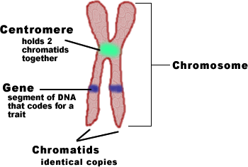
What you are Replicating and dividing - a close look at a chromosome

Regulation
- Cyclins are proteins which regulate the cell cycle. There are many types.
- CDK (Cyclin Dependent Kinase) along with cyclins are major control switches for the cell cycle causing cell to move from G1 to G2
- MPK (Maturation Promoting Factor) inclueds the CDK and cyclins that triggers progession through the cell cycle.
- p53 is a protein that functions to block the cell cycle if DNA is damaged. If the damage is severe, it can cause apoptosis (cell death).
- p27 is a protein that binds to cyclin and cdk blocking entry to S phase.
Confusing "C" words that all sound the same but mean different things in the cell cycle:
Chromatin: Unwound DNA in nucleus, used to make mRNA later. Used in chromosomes.
Centomere: The middle point in a chromosome.
Centrosome: 2 centrioles.
Centrioles: The "T" shaped structures that pull that stretch the cell during mitosis.
Chromatid: One half (a ">" or "<" shape) of a chromosome. There are 2 sister chromatids for a chromosome.
Chromosome: Tightly wound DNA that contains histones, or proteins and is made up of chromatin. Chromosomes have 2 identical Chromatids that form an "X" shape. When these chromatids seperate at anaphase, they become their own full fledged chromosome.
10.1 Cell Growth
· the larger a cell becomes, the more demands the cell places on its DNA and the more trouble the cell has movie enough nutrients and wastes across the cell membrane
· cell division- the process by which a cell divides into two new daughter cells
· As the length of a cell increases, its volume increases faster than its surface area. the resulting decrease in the cell’s ratio of surface area to volume makes it more difficult for the cell to move needed materials in and waste products out.
10.3 Regulating the Cell Cycle
· cyclin- one of a family of closely related proteins that regulate the cell cycle in eukaryotic cells
· cancer- disorder in which some of the body’s own cells lose the ability to control growth. A class of diseases in which a group of cells lose the capacity to control all cell growth, invasion, and metastasis.
· Cyclins regulate the timing of the cell cycle in eukaryotic cells.
· Cancer cells do not respond to the signals that regulate the growth of most cells. As a result, they form masses of cells called tumors that can damage the surrounding tissues. These cells are also able to travel to different parts of the body, and affect those areas negatively, mostly in the formation of other tumors.

---This video shows an overview of the cell cycle with regards to G1, G0, S, M, and G2.
Here is a detailed diagram of the Cell Cycle, showing the progression as well as duration of each step:

Description of Phases:
Resting (G0 phase)
The term "post-mitotic" is sometimes used to refer to both quiescent and senescent cells. Nonproliferative cells in multicellular eukaryotes generally enter the quiescent G0 state from G1 and may remain quiescent for long periods of time, possibly indefinitely (as is often the case for neurons). This is very common for cells that are fully differentiated. Cellular senescence is a state that occurs in response to DNA damage or degradation that would make a cell's progeny nonviable; it is often a biochemical alternative to the self-destruction of such a damaged cell by apoptosis.
Interphase
G1 phase
The first phase within interphase, from the end of the previous M phase until the beginning of DNA synthesis is called G1 (G indicating gap). It is also called the growth phase. During this phase the biosynthetic activities of the cell, which had been considerably slowed down during M phase, resume at a high rate. This phase is marked by synthesis of various enzymes that are required in S phase, mainly those needed for DNA replication. Duration of G1 is highly variable, even among different cells of the same species.
S phase
The ensuing S phase starts when DNA synthesis commences; when it is complete, all of the chromosomes have been replicated, i.e., each chromosome has two (sister) chromatids. Thus, during this phase, the amount of DNA in the cell has effectively doubled, though the ploidy of the cell remains the same. Rates of RNA transcription and protein synthesis are very low during this phase. An exception to this is histone production, most of which occurs during the S phase
This picture is of the first two phases of the cell cycle, G1 and S.

G2 phase
The cell then enters the G2 phase, which lasts until the cell enters mitosis. Again, significant protein synthesis occurs during this phase, mainly involving the production of microtubules, which are required during the process of mitosis. Inhibition of protein synthesis during G2 phase prevents the cell from undergoing mitosis.
This picture is a picture of the G2 Phase.

<object width="560" height="340"><param name="movie" value="http://www.youtube-nocookie.com/v/DUKwBQlJipM&hl=en_US&fs=1&rel=0"></param><param name="allowFullScreen" value="true"></param><param name="allowscriptaccess" value="always"></param><embed src="http://www.youtube-nocookie.com/v/DUKwBQlJipM&hl=en_US&fs=1&rel=0" type="application/x-shockwave-flash" allowscriptaccess="always" allowfullscreen="true" width="560" height="340"></embed></object>
The professor in the above video does a good job explaining the processes of interphase. While it is often overlooked as a slow-paced resting period during the cell cycle, interphase is actually vital to the process as this is where the cell grows and prepares for the division by replicating its DNA.
Mitosis (M Phase)
Main article: Mitosis
The relatively brief M phase consists of nuclear division (karyokinesis) and cytoplasmic division (cytokinesis). In plants and algae, cytokinesis is accompanied by the formation of a new cell wall. The M phase has been broken down into several distinct phases, sequentially known as:
- prophase (This is the longest period of the complete cell cycle during which DNA replicates, the centrioles divide, and proteins are actively produced)
- prometaphase (In this stage the nuclear envelope breaks down so there is no longer a recognizable nucleus)
- metaphase (Tension applied by the spindle fibers aligns all chromosomes in one plane at the center of the cell)
- anaphase (Spindle fibers shorten, the kinetochores separate, and the chromatids (daughter chromosomes) are pulled apart and begin moving to the cell poles)
- telophase (The daughter chromosomes arrive at the poles and the spindle fibers that have pulled them apart disappear)
- cytokinesis (The spindle fibers not attached to chromosomes begin breaking down until only that portion of overlap is left. It is in this region that a contractile ring cleaves the cell into two daughter cells. Microtubules then reorganize into a new cytoskeleton for the return to interphase)
Mitosis is the process in which a eukaryotic cell separates the chromosomes in its cell nucleus into two identical sets in two daughter nuclei.It is generally followed immediately by cytokinesis, which divides the nuclei, cytoplasm, organelles and cell membrane into two daughter cells containing roughly equal shares of these cellular components. Mitosis and cytokinesis together define the mitotic (M) phase of the cell cycle - the division of the mother cell into two daughter cells, genetically identical to each other and to their parent cell.
Mitosis occurs exclusively in eukaryotic cells, but occurs in different ways in different species. For example, animals undergo an "open" mitosis, where the nuclear envelope breaks down before the chromosomes separate, while fungi such as Aspergillus nidulans and Saccharomyces cerevisiae (yeast) undergo a "closed" mitosis, where chromosomes divide within an intact cell nucleus. Prokaryotic cells, which lack a nucleus, divide by a process called binary fission.
The process of mitosis is complex and highly regulated. The sequence of events is divided into phases, corresponding to the completion of one set of activities and the start of the next. These stages are prophase, prometaphase, metaphase, anaphase and telophase. During the process of mitosis the pairs of chromosomes condense and attach to fibers that pull the sister chromatids to opposite sides of the cell. The cell then divides in cytokinesis, to produce two identical daughter cells.
Because cytokinesis usually occurs in conjunction with mitosis, "mitosis" is often used interchangeably with "mitotic phase". However, there are many cells where mitosis and cytokinesis occur separately, forming single cells with multiple nuclei. This occurs most notably among the fungi and slime moulds, but is found in various different groups. Even in animals, cytokinesis and mitosis may occur independently, for instance during certain stages of fruit fly embryonic development. Errors in mitosis can either kill a cell through apoptosis or cause mutations that may lead to cancer.
This picture outlines the 5 main steps of Mitosis, however it excludes the first, the Interphase.

 This is a video that also demonstrates the mitosis process that has been described. It's extremely helpful and gives you a view right inside the cell so that you can get a clear look and understanding of the 5 main steps in mitosis.

This is a creative and fun video that helped me learn more about how mitosis takes place and what it does.

This diagram shows where the p53 and p27 come into play during the cwell cycle.
Cell Cycle in Cancer
The cell cycle, the process by which cells progress and divide, lies at the heart of cancer. In normal cells, the cell cycle is controlled by a complex series of signaling pathways by which a cell grows, replicates its DNA and divides. This process also includes mechanisms to ensure errors are corrected, and if not, the cells commit suicide (apoptosis). In cancer, as a result of genetic mutations, this regulatory process malfunctions, resulting in uncontrolled cell proliferation.
Cyclacel�s drug discovery and development programs build on recent scientific advances in understanding these molecular mechanisms. Through our expertise, we are developing cell cycle-based, mechanism-targeted cancer therapies that emulate the body�s natural process in order to stop the growth of cancer cells. This approach can limit the damage to normal cells and the accompanying side effects caused by conventional chemotherapeutic agents.
Additional Information
The cell cycle involves a complex series of molecular and biochemical signaling pathways. As illustrated in the diagram above the cell cycle has four phases:
- the G1, or gap, phase, in which the cell grows and prepares to synthesize DNA;
- the S, or synthesis, phase, in which the cell synthesizes DNA;
- the G2, or second gap, phase, in which the cell prepares to divide; and
- the M, or mitosis, phase, in which cell division occurs.
http://www.biology.arizona.edu/Cell_Bio/tutorials/cell_cycle/main.html
This website helps give additional information about mitosis and the cell cycle in dumbed down terms.. check it out
Cancer and the Cell Cycle
Cancer cells have mutations in the genes that control the cell cycle. Some of these genes tell the cell cycle to proceed, in other words they are "go" genes like the accelerator in your car. These are called proto-oncogenes. Some of these genes tell the cell cycle to stop when the cell is done dividing and no more cells are needed. These are called tumor suppressor genes. They are like the brake on your car. If either of these are mutated, it causes the cell cycle to go too fast, like having your accelerator stuck to the floor or losing your brakes. So the cancer cells divide too fast and pile up in one area, this is called a tumor. This is an extremely complex issue. Essentially cell cycle is everythingwhen it comes to control of growth in cells and that is just what cancer is...a loss in the control of cell growth. There are many known and probably far too many more unknown control points in the cell cycle that normal cells lose when the become cancerous. These can be in any of thestages, G1, G2, or even S. There are a number of genes which exert control in the cell cycle, on eof the best known is P53 which is implicated in over 50% of human cancers.

 Here is a youtube video that shows how cancer develops in the cell cycle.
Here is a youtube that shows how stem cells could kill cancer cells.

Origins of cancer
All cancers begin in cells, the body's basic unit of life. To understand cancer, it's helpful to know what happens when normal cells become cancer cells.
The body is made up of many types of cells. These cells grow and divide in a controlled way to produce more cells as they are needed to keep the body healthy. When cells become old or damaged, they die and are replaced with new cells.
However, sometimes this orderly process goes wrong. The genetic material (DNA) of a cell can become damaged or changed, producing mutations that affect normal cell growth and division. When this happens, cells do not die when they should and new cells form when the body does not need them. The extra cells may form a mass of tissue called a tumor.

Not all tumors are cancerous; tumors can be benign or malignant.
- Benign tumors aren't cancerous. They can often be removed, and, in most cases, they do not come back. Cells in benign tumors do not spread to other parts of the body.
- Malignant tumors are cancerous. Cells in these tumors can invade nearby tissues and spread to other parts of the body. The spread of cancer from one part of the body to another is called metastasis
Metastasis occurs when the cancer cells break away or leak and spill away from the tumor. They then enter blood vessels and once they get into there they can work their throughout the body and be deposited within normal tissue. The most common places for them to metastisis is in the lungs, liver, brain, and bones. Different types of cancers have their tendencys to metastisize in certain places such as prostate cancer, which tipically metastisizes to bones. The cells of the metastisized tumor resemble those of the original.
The following link displays an animation on how cell regulators are able to prevent the creation or spread of cancerous cells. This example focuses primarily on p53, but other regulators include Cdk and p27.
http://www.hhmi.org/biointeractive/media/using_p53-lg.mov
STUDY HELP
http://www.quia.com/quiz/869768.html
This link quizes you on the cell cycle and I found it very helpful.
http://www.biology.arizona.edu/Cell_Bio/tutorials/cell_cycle/main.html
this also is a page where you can get a better understanding of the cell cycle and mitosis
The pie chart below shows the time spent in the 4 stages of the cell cycle as the majority of time spent in the cell cylce is in the the first three stages which equates to interphase. the fourth that contains the M of the cell cycle contains prophase through telophase which is mitotic phase.

The cell cycle is a cylce because it doesn't stop, it just repeats its self. The cell does its normal activities, and then it divides. Once it has divided it does its normal activities again because it is one cell, until cyclin tells it that it needs to duplicate again. This cylce however does end when the cell dies because of DNA that is too short, a mutation, or just because it has ran its course.

Here is a very simple diagram of the cell cycle, showing the phases go from g1 to S to g2 and then to Mitosis.
http://highered.mcgraw-hill.com/sites/0072495855/student_view0/chapter2/animation__how_the_cell_cycle_works.html
^Copy and paste into URL
This website is a very good review of how the cell cycle works. It's an easy understanding video and will definitely help you review.

this picture shows the cell cycle in good detail with good labels.

This photo is a great representation of how the cell cycle works and it's stages. (it goes top row, left to right, bottom row left to right)

This video is made by two students so it is kind of funny..but still helpful! It gives a good explanation of the G1 phase, S phase, and G2 phase.
Comments (0)
You don't have permission to comment on this page.